
Authors

Catheter ablation for atrial fibrillation is a well-known corrective procedure. Thanks to new innovative three-dimensional (3D) mapping systems, more cardiac arrhythmia patients can now benefit from zero radiation exposure during transcatheter procedures.
Pioneered by Professor Sabine Ernst, consultant cardiologist and reader in cardiology at Royal Brompton and Harefield hospitals, the unique 3D mapping technique removes the need for radiation.
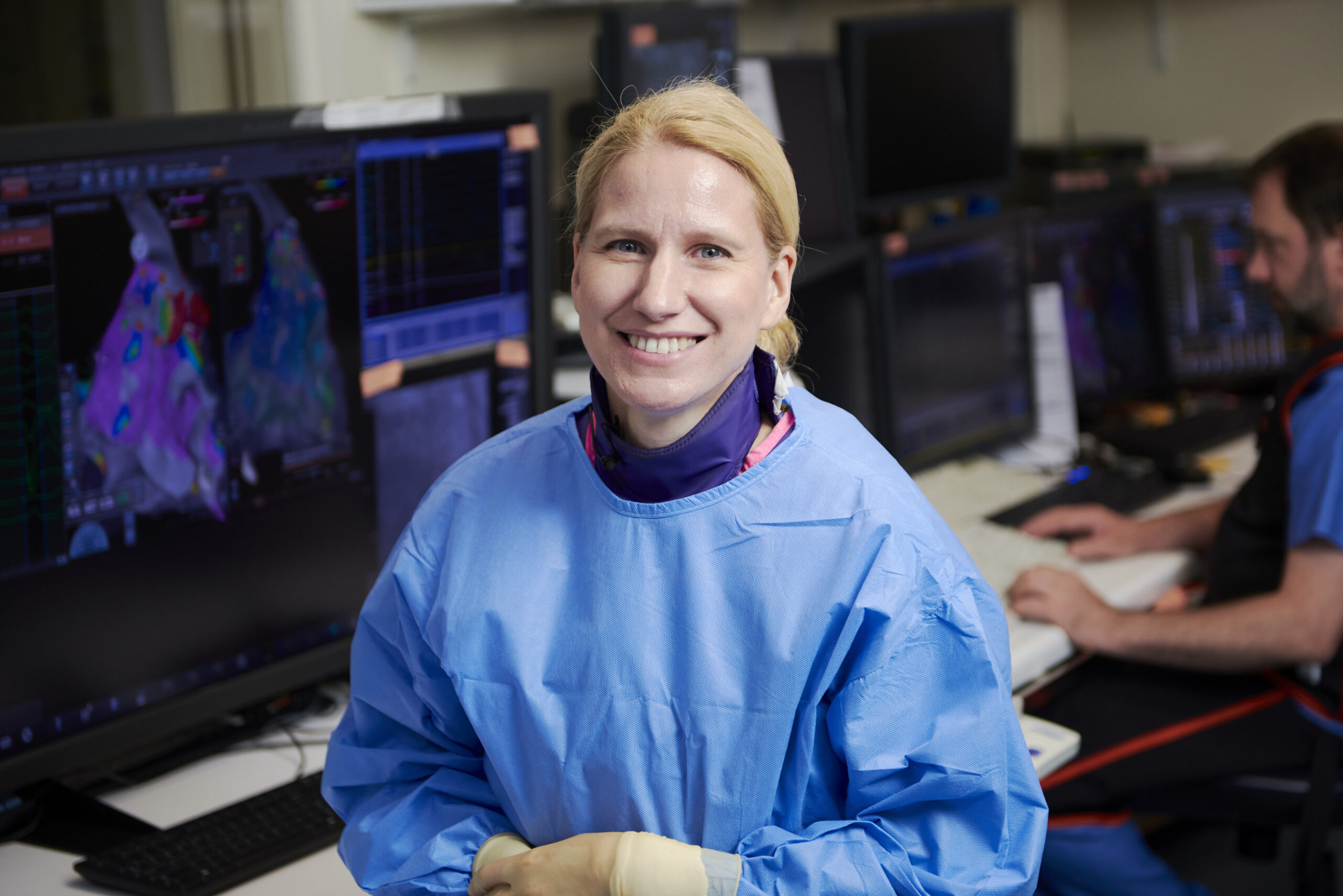
Pioneered by Professor Sabine Ernst, the unique 3D mapping technique removes the need for radiation
The non-fluoroscopic catheter ablation for atrial fibrillation technique is an innovative treatment only available at the Royal Brompton Hospital. Professor Ernst is, so far, the only practitioner using this approach which entirely avoids using x-rays during the invasive procedure.
Arrhythmias, or abnormal heart rhythms, are widely experienced by people across the world. Atrial fibrillation is the most common form of arrhythmia and is a major cause of strokes. During Arab Health 2019, the biggest medical conference in the Middle East, Dr Ernst will showcase this unique approach during a simulated surgical presentation.
A US study featured in The Lancet revealed the prevalence of atrial fibrillation in the Middle East triples with each decade of life compared to other developed countries, where it doubles with each decade of life. Additionally, twice as many women are affected by mitral stenosis than men in the Middle East, which leads them to develop atrial fibrillation more often. These trends may be attributed to increases in sedentary lifestyles, ageing, obesity, diabetes, and hypertension. This suggests atrial fibrillation and stroke will become increasingly important health issues in the Middle East.
Atrial fibrillation causes the heart to beat irregularly and faster than normal and, if medication hasn’t worked, the usual procedure for rectification is catheter ablation. This involves correcting abnormal electrical impulses in the heart, using x-rays in combination with 3D mapping to navigate the catheter.
The downside of the conventional ablation procedure is the exposure to radiation through x-rays, as they are linked to an increased risk of developing cancer in the future. This is particularly relevant when a patient is young, female and of child-bearing age or requires many x-ray guided procedures.
A different roadmap
Professor Ernst explains, “if the patient’s heart is otherwise healthy, catheter ablation of atrial fibrillation works with the majority of cases with one treatment. But if a patient has heart disease or the atrial fibrillation is already persistent, then the procedure is less likely to work the first time and a patient may need multiple ablations. This automatically increases their radiation exposure.”
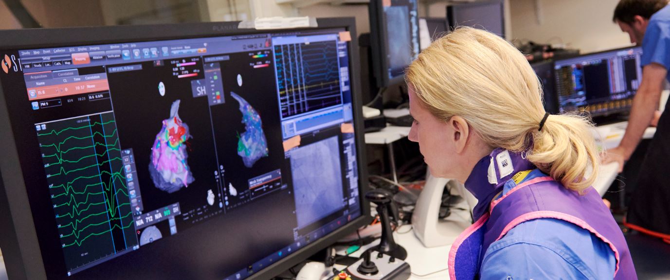
To reduce patients’ exposure to radiation, Professor Ernst has developed a new structured approach to catheter ablation of atrial fibrillation. This replaces x-ray navigation with an electro-anatomical mapping system that creates 3D images showing the catheters within the heart during the procedure. It also displays the transseptal needle used to enter the left side of the patient’s heart.
In addition, Professor Ernst uses 3D roadmaps from either cardiac magnetic resonance imaging (CMR) or computed tomography (CT) scans. These 3D roadmaps show the detailed anatomy of the patient’s heart and help to locate the catheters during the procedure. The mapping system uses a special catheter equipped with a sensor to allow exact localisation.
Zero or minimal radiation
With 3D mapping, the total radiation time from x-rays can be substantially reduced or completely eliminated. For a growing number of patients who need to undergo an ablation, the exposure to x-ray radiation can be zero, or reduced to just a few seconds.
With this approach, the patient’s lifetime ‘radiation bill’ is not added to with potentially harmful radiation, therefore the cancer risk is not increased by the procedure or procedures.
Some patients, however, do need to undergo x-rays for their treatment, for example, those with implanted devices such as pacemakers, defibrillators (ICDs) and biventricular devices. Depending on the position of an artificial heart valve, patients with a metallic heart valve may also require navigation using x-rays.
Since 2013, Professor Ernst has carried out over 60 catheter ablations without using any radiation at all.
How catheter ablation with 3D mapping works
Professor Ernst starts by looking at a 3D image of the patient’s heart, gained through CMR imaging. This removes the need for radiation unless a patient has a contraindication with CMR and requires a CT instead.
To begin the procedure, a small puncture in the groin is made with a special needle which advances the catheter towards the heart, guided by images provided by the 3D mapping system.
Professor Ernst explains: “In the left atrium, I treat the entry points, the so-called pulmonary veins, and this reverts the heart to its normal rhythm. Typically, I also perform an ablation in the right atrium for the best outcome.”
The procedure has equal success rates whether using x-ray or 3D mapping systems for navigation, and the approach doesn’t increase a patient’s cumulative lifetime exposure to x-rays.
In both cases, Professor Ernst says the whole procedure takes between an hour and a half and two hours. After the patient comes out from under the general anaesthetic, they need six hours’ bed rest and a stay overnight on the ward. The next day, the team conducts safety checks, including an echocardiogram, and the patient is free to go home. Patients can return to normal physical activity after 10 days.
Benefits for patients
The main advantage for patients is a reduction in their x-ray exposure. Professor Ernst says: “It’s cumulative – every time a patient has a scan they up their risk. Anything to reduce this exposure is good for them.”
This benefit is particularly important for young women and all female patients of childbearing age. In addition, catheter ablation using zero or minimal radiation is a minimally invasive process which is an attractive option for females.
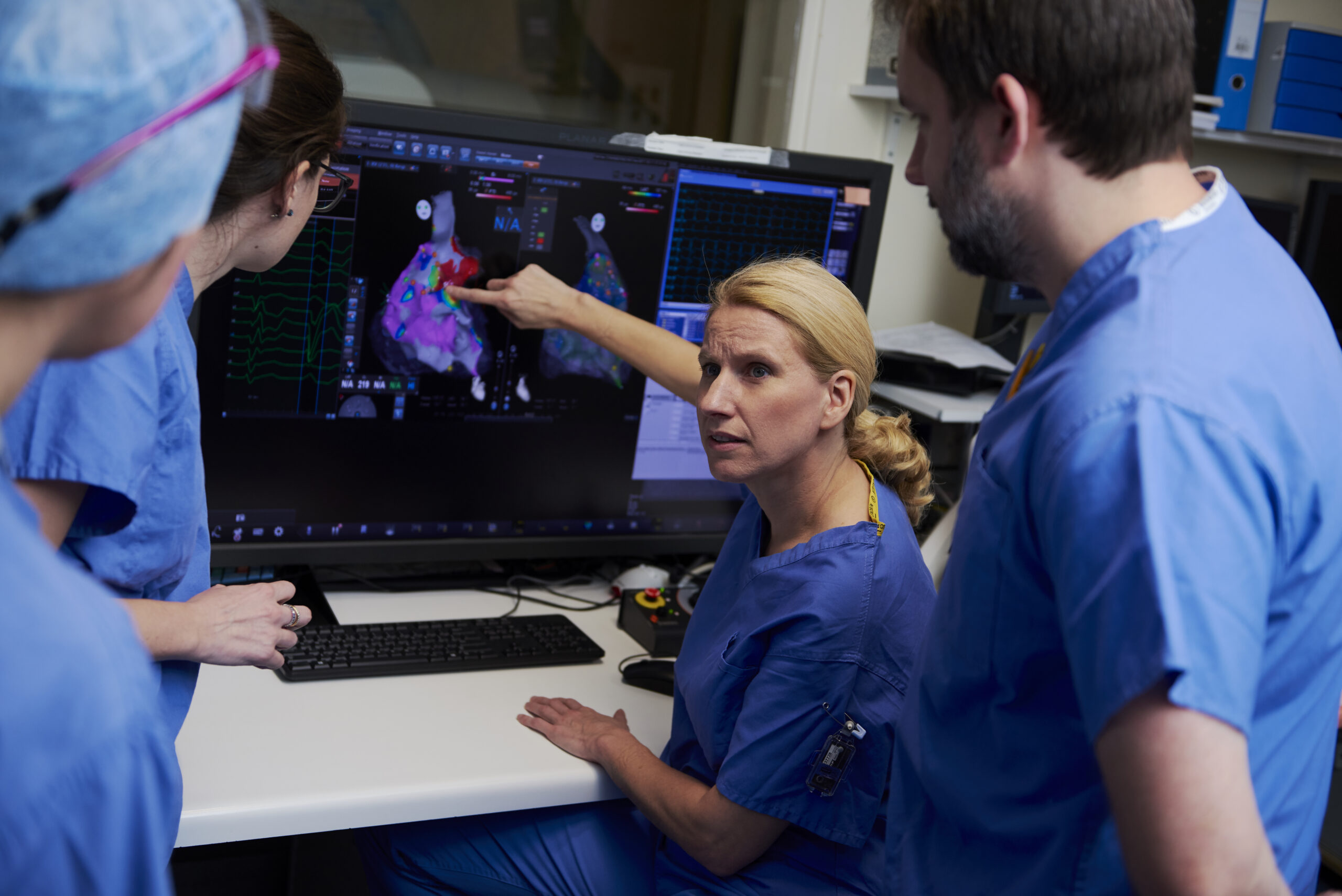
Professor Ernst has a strong appeal to women who prefer a female consultant in the event they need treatment for a cardiac arrhythmia such as atrial fibrillation. Professor Ernst can also arrange for an all-female team – comprising an anaesthetist, two nurses and two technicians – to work alongside her on the catheter ablation.
Links with the Middle East
Guy’s and St Thomas’ Specialist Care has a strong and long established relationship with the Gulf region. The organisation works closely with health authorities including the Dubai Health Authority, Dubai Ministry of Health and Prevention, and Hamad Medical Corporation, Qatar.
Known across the world over for our expertise, standard of care and research success in lung and heart disease, we operate a visiting doctor programme with key hospitals across the Middle East region. The programme helps to provide better clinical outcomes and strengthen relationships with the region’s healthcare providers.
Professor Ernst says: “The programmes and collaborations in place between Guy’s and St Thomas’ Specialist Care and hospitals in the Middle East region are there to ensure patients receive the best care while visiting the UK for treatment. Crucially, we also look to ensure that continuation of care is transferred back to the country. By sharing our knowledge, patients receive excellent care wherever they – this makes a real difference.”
Professor Ernst also plans to cascade specific knowledge of her 3D mapping approach to catheter ablation across London and Europe in 2019. In the future, she is hopeful that this innovative and beneficial approach will be available globally.
At a glance
Procedure
Zero radiation catheter ablation using structured 3D mapping
Carried out by
Professor Sabine Ernst
Consultant cardiologist
Reader in cardiology at Imperial College London
What problem does it solve?
It allows many arrhythmias to be treated with zero radiation exposure from x-rays. In most cases, a structured 3D mapping approach doesn’t use radiation, so it doesn’t increase a patient’s cumulative lifetime exposure to x-rays (which can increase the risk of developing cancer later in life).
How does it work?
3D mapping helps to find the exact place in a patient’s heart responsible for a faulty electrical pathway. During the ablation procedure, this site can be treated to stop the arrhythmia.