
Authors

About 1% of babies born worldwide have congenital heart disease (CHD), a term which refers to range of heart defects that develop in the womb. Risk factors of CHD include having a family history of heart problems, maternal diabetes and advanced maternal or paternal age. Unfortunately, many babies are not diagnosed with a heart problem until after they are born. However, our consultant fetal cardiologists at Evelina London Children’s Hospital have developed ways to detect heart problems in the fetus before birth, allowing for better management of the baby after birth.
Fetal diagnosis for better outcomes in later life
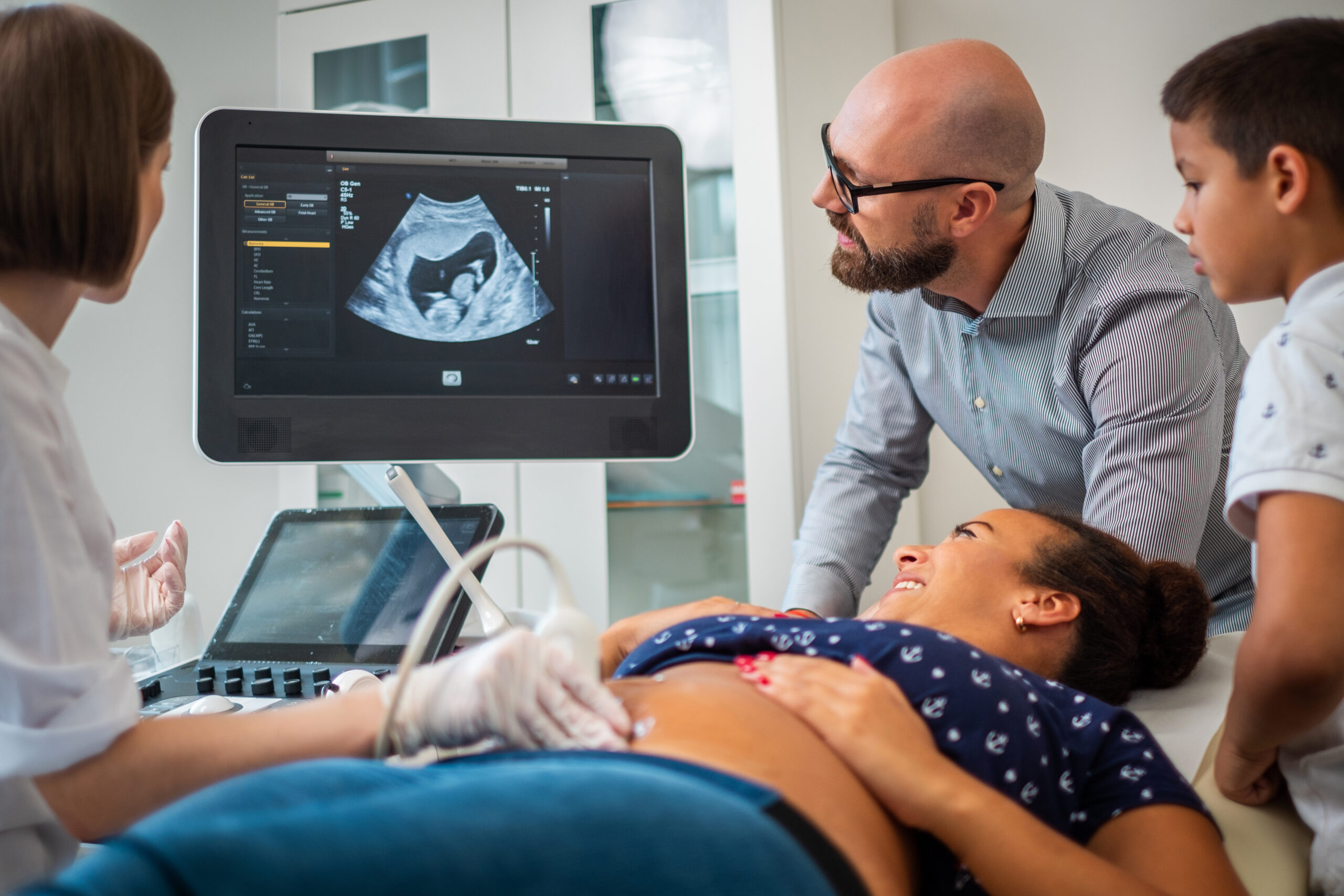
Every pregnant woman should be offered an ultrasound scan at 18 to 20 weeks, which is designed to check that all organs of the fetus, including the heart, are developing properly.
Dr Owen Miller, consultant in paediatric and fetal cardiology at Evelina London, part of Guy’s and St Thomas’ NHS Foundation Trust, explains: “Many women don’t find out their baby has a heart problem until after they are born. It is important to precisely diagnose the type of congenital heart disease in the unborn baby during pregnancy. If the infant requires surgical intervention soon after delivery, we can prepare for surgery and achieve the best outcomes,” explains Dr Miller.
Artificial intelligence to help identify heart disease
Our consultant fetal cardiologists at Evelina London and at our university academic partner Kings College London have developed “machine learning” technology, which helps detect irregularities in the heart structure, which may indicate a heart problem in the baby.
To “train” the machine, our consultants pass thousands of heart ultrasound images through a computerised network multiple times, and the “machine learning” gets better each time at telling the difference between images. When the network identifies a heart with a different appearance, our consultants can review the scan in further specific detail.
While the technology does not remove the need for human review, “using artificial intelligence offers a quicker way to scan for heart irregularities. We can save seven minutes when compared to a standard, manual scan,” says Dr Thomas Day, paediatric and fetal cardiologist at Evelina London.
3D MRI technology for multi-angled views
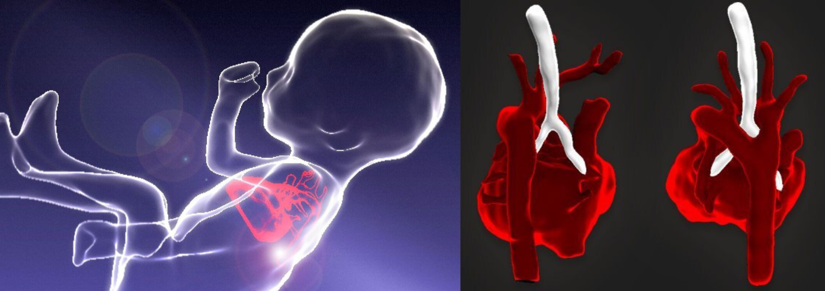
The technique transforms standard images into clear three-dimensional images (left). A 3D MRI scan of a fetal heart (right).
Cardiac Magnetic Resonance Imaging (MRI) is a safe and well-established method for viewing structures of the heart in babies and young children. However, due to the small size of the heart before birth and the constant movement of the baby, “standard” MRI scans can be extremely unreliable, with only a few centres around the world offering this technology.
To overcome these challenges, our teams at Evelina London, St Thomas’ Hospital and King’s College London were the first in the world to develop accurate and reliable fetal cardiac MRI, using a new motion correction technique which involves taking multiple images with the MRI scan and stacking them up so that we can reconstruct the fetal heart in three dimensions. Pushing the technology a step further, these 3D images can then be converted into a physical representation of the heart, via 3D printing technology.
“Being able to show a parent a 3D version of their unborn baby’s heart is very helpful, and this technology allows us to create images of the heart in a way that the clinicians and patients can better understand,” says Dr Kuberan Pushparajah, consultant cardiologist at Evelina London. “We were the first ones to be able to do this in a systematic, valid and reproducible way,” he adds.
“When a patient comes for a heart MRI scan, they will also receive detailed imaging of the baby’s brain and body, all of which are performed and reported by world-leading experts in this type of imaging. Along with our experienced colleagues in fetal medicine and obstetrics, this allows us to provide the most comprehensive care for babies and their families,” says Dr David Lloyd, consultant paediatric and fetal cardiologist at Evelina London.
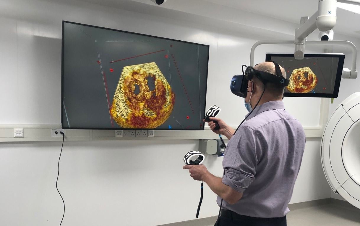
Prof John Simpson using VR technology to view images of the heart
Furthermore, our consultants have developed a way to transfer the 3D images into a virtual reality (VR) environment. Equipped with a VR headset and handset controls, the technology allows our cardiologists and surgeons to become immersed into the images as if they are viewing a real 3D beating heart.
These 3D images can then be shared among our experts, allowing for second opinions and improving shared understanding of the baby’s heart. Using this technology, we are able to examine and investigate how the heart may respond to different forms of treatment.
Professor John Simpson, professor of paediatric and fetal cardiology at Evelina London, comments: “This is cutting-edge technology and is something that could give us a huge amount of additional, dynamic information.”
Get in touch
If you are concerned about the health of your unborn baby’s heart and would like to speak to one of our fetal cardiology specialists, please contact our team to arrange an appointment.
Related content
-
Antenatal tests
As part of our leading private maternity service we offer a range of blood tests, swabs and screenings to support you during your pregnancy.
-
Fetal cardiology
As one of the largest fetal cardiology units in Europe we manage heart problems in babies before birth.
-
Revolutionary technology produces three-dimensional fetal cardiac images of the heart before birth